


Imaging an Entire Artery by determining the exact location and the orientation of the ultrasound transducer (#2018017)
Chen Speter, MD , Doron Manzur, MD , Chen Giladi, Dr.
Contact Us for more information:Tel Hashomer Medical Research, Infrastructure and Services Ltd.Innovation.office@sheba.health.gov.il |
| Categories | Ultrasound, Sonography, Ultra-sonography, Arterial Imaging |
| Development Stage | Initial prototype |
| Patent Status | pending |
Background
Diseases of peripheral arterial system are one of the common causes of limb pain, especially in elderly patients. Intermittent claudication due to peripheral arterial disease occurs in 3 to 6% of elderly population. Clinical evaluation followed by Doppler ultrasound and CT angiogram gives the definitive diagnosis. After the advancement of CT techniques, it is possible to cover entire limbs within a few seconds. Optimization of intravenous contrast with blood flow and CT scanning gives higher spatial resolution and coverage of more than 120cm. Computers with graphics project images in 3D, volume rendered images and curved planar images. Imaging of the entire arterial system is possible using CT angiography and has found excellent concordance with digital subtraction imaging.
As there is need of accurate, reliable and reproducible non-invasive evaluation, in a point of care set-up, we are developing a novel imaging device that collects US images (with the additional layers of information gained by the Doppler) and fuses them to one whole organ. The results are a 4D image, using the same philosophy as the CT: information about the specific coordinates for each image allows the computer to create the tomography of the object.
The Technology and Innovation
The novelty in this invention is related to the ability to collect precise probe positions and orientations. The images are then being processed with two innovative algorithms, and output the computed result in a new visualization fused 4D whole organ image. Our PROOF OF CONCEPT reveals that the position of the probe is exceptionally accurate and is achieved with a very low-cost apparatus.

The Benefits
- For the same quality of precise imaging as the CT, the device avoids radiation and contrast medium.
- Using Duplex gives not only information about the anatomy of the artery, but also data about the velocity and the capacity of the blood flow.
- Our device allows inexperienced users to operate the US imaging system and perform a high-quality examination of a patient, thus reducing to a minimum the dependence of the operator.
- Our device and algorithms are capable of producing much more accurate segmentation and better resolution by eliminating the different noise sources, which are the downsides of the US.
- The final computed whole organ is unique and informative and allows registration with other imaging modalities (CTA, angiogram, MRA).
Although our device can be utilized in any compatible US organ scanning procedure (e.g., liver, gallbladder, thyroid, etc.), we think that the best-added value would be in the vascular imaging field, since it can also collect blood velocity and capacity (Doppler data). This data is crucial for surgical decisions, making this tool a 4D imaging device that can be anatomically precise as a CT, but with the advantage of the physiological vascular information.
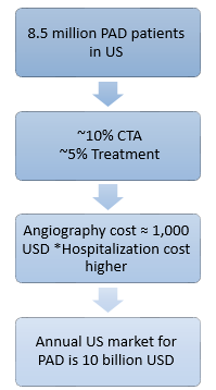
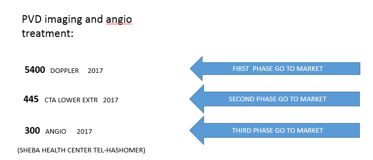
The Market
The Team
- Dr. Giladi is a physicist with expertise in machine learning, signal processing, and robotics.
- Dr. Manzor MD. has experience with several medical startups, currently working as a clinical development manager at "Novocure," a medical device company.
- Dr. Speter MD. is a board-certified general surgeon and a board-certified vascular surgeon. Dr. Speter has been working at Sheba hospital for the past 12 years.
Contact Us for more information:Tel Hashomer Medical Research, Infrastructure and Services Ltd.Innovation.office@sheba.health.gov.il |